Home » Without Label » Bones Of Female Back - Image of female skeleton stock illustration. Image of ... : Diagram of a human female skeleton, back view.
Bones Of Female Back - Image of female skeleton stock illustration. Image of ... : Diagram of a human female skeleton, back view.
Bones Of Female Back - Image of female skeleton stock illustration. Image of ... : Diagram of a human female skeleton, back view.. The spinal cord has a varying width, ranging from 13 mm (1/2 in) thick in the cervical and lumbar regions to 6.4 mm (1/4 in) thick in the thoracic area. Whilst each section of the vertebral column consists of. The axial skeleton is made up of the skull, backbone, breastbone, and ribs. Without the cushioning provided by the cartilage, your bones may rub together, causing pain. The risk for osteoporosis and osteopenia—low bone density that's not yet in the osteoporosis range—is higher in women because female bones typically are smaller and less dense than male bones.
See female skeleton stock video clips. Chronically low muscle mass is associated with low bone mass. There are two hip bones, one on the left side of the body and the other on the right. Browse 222 lower back skeleton stock photos and images available, or start a new search to explore more stock photos and images. Diagram of a human female skeleton, back view.
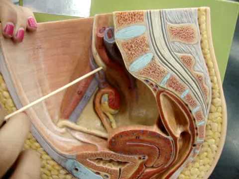
Female Pelvis Model (Exam #3) - YouTube from i.ytimg.com But men are also at risk. Muscle anatomy triceps 12 photos of the muscle anatomy triceps. Diagram of a human female skeleton, back view. Women have a wider pelvis (hips) than men because the opening has to be wide enough to let a baby pass through when it's born. Roughly 500,000 spinal fractures occur due to osteoporosis every year. Together, they form the part of the pelvis called the pelvic girdle. Bones has been gone for a while now, and its cast members have moved on to other things, professionally and personally. The bones of the superior portion of the skull are known as the cranium and protect the brain from damage.
Together, they form the part of the pelvis called the pelvic girdle.
Bones make up about 14 percent of our body weight. Flexibility especially in the lower back and neck allowing us to bend and twist in a full variety of movements strength provided by the bones discs joints and supportive muscles and connective tissue that allows us to stand upright and move about with precision. Women have a wider pelvis (hips) than men because the opening has to be wide enough to let a baby pass through when it's born. Spinal osteoarthritis causes a breakdown of the fibrous cartilage in the facet joints. Because bone growth naturally slows down later in life, osteoporosis can affect both men and women over age 50. See human back anatomy stock video clips. Osteoporosis is one of the most common issues leading to back pain in the u.s. An image shared on social media has shown a bulge in a woman's back where he bones have moved to make way for her baby during. Browse 222 lower back skeleton stock photos and images available, or start a new search to explore more stock photos and images. Everything else that hangs from this, like the arms, legs, shoulders, and hips, is called the appendicular skeleton. Some causes are specific to women, like pms or endometriosis, while other causes can happen to anyone. Together, they form the part of the pelvis called the pelvic girdle. The risk for osteoporosis and osteopenia—low bone density that's not yet in the osteoporosis range—is higher in women because female bones typically are smaller and less dense than male bones.
Lower back pain in women can be caused by many different conditions and underlying factors. All evidence points to the victim being booth. Together, they form the part of the pelvis called the pelvic girdle. Bones make up about 14 percent of our body weight. The bones of the back, together, make up the vertebral column.the vertebral column is made up of 5 sections:
Silkie Chicken's Black Skin And Bones Thought To Aid ... from images.medicaldaily.com Temperance bones brennan portrayed by emily deschanel. Woman's bones in back move during childbirth in astonishing photo. Together, they form the part of the pelvis called the pelvic girdle. All evidence points to the victim being booth. Without the cushioning provided by the cartilage, your bones may rub together, causing pain. The second cause is a loss of bone once a person has reached peak bone mass. The risk for osteoporosis and osteopenia—low bone density that's not yet in the osteoporosis range—is higher in women because female bones typically are smaller and less dense than male bones. Peak bone mass is typically reached by age 20 in men and age 30 in women.
The human spinal cord, part of the central nervous system, is around 45 cm (18 in) in men and around 43 cm (17 in) long in women.
It is made up of five fused vertebral bones. The bones of the back, together, make up the vertebral column.the vertebral column is made up of 5 sections: Human body anatomy female female anatomy muscle shoulder blade pain anatomy back muscles bones man female anatomy body muscles in a body female anatomy muscole shoulder concept muscular sysyem. Together, they form the part of the pelvis called the pelvic girdle. The bones that make up your spine (vertebrae) can weaken to the point of collapsing, which can result in back pain, lost height and a hunched forward posture. Some causes are specific to women, like pms or endometriosis, while other causes can happen to anyone. The bones of the superior portion of the skull are known as the cranium and protect the brain from damage. Diagram of a human female skeleton, back view. Chronically low muscle mass is associated with low bone mass. 9 photos of the diagram of female back muscles. Here's a look into what became of the actors who portrayed all those. Women have a wider pelvis (hips) than men because the opening has to be wide enough to let a baby pass through when it's born. Even stronger grip strength and stronger back muscles are associated with higher bone density.
It runs down the centre of the body. Xray of hip skeleton health xray pelvis full skeleton joint & muscles chiropractor woman bones care bones and joints hand female body xray exercise and bone health. But men are also at risk. Bones of female back : The first is a failure to reach one's peak bone mass, which is the maximum amount of bone a person is genetically programmed to build in her lifetime.
Bony Pelvis Anatomy | Bone and Spine from boneandspine.com The red lines point individual bones and the names are writen in singular, the blue lines conect to group of bones and are in plural form. Because bone growth naturally slows down later in life, osteoporosis can affect both men and women over age 50. All evidence points to the victim being booth. The thigh bone (femur) is the longest bone in the body. The bones of the pelvis and lower back work together to support the body's weight, anchor the abdominal and hip muscles, and protect the delicate vital organs of the vertebral and abdominopelvic cavities. Human body anatomy female female anatomy muscle shoulder blade pain anatomy back muscles bones man female anatomy body muscles in a body female anatomy muscole shoulder concept muscular sysyem. An image shared on social media has shown a bulge in a woman's back where he bones have moved to make way for her baby during. The second cause is a loss of bone once a person has reached peak bone mass.
The spinal cord has a varying width, ranging from 13 mm (1/2 in) thick in the cervical and lumbar regions to 6.4 mm (1/4 in) thick in the thoracic area.
The risk for osteoporosis and osteopenia—low bone density that's not yet in the osteoporosis range—is higher in women because female bones typically are smaller and less dense than male bones. Here's a look into what became of the actors who portrayed all those. Spinal osteoarthritis causes a breakdown of the fibrous cartilage in the facet joints. It is made up of five fused vertebral bones. Women have a wider pelvis (hips) than men because the opening has to be wide enough to let a baby pass through when it's born. Diagram of a human female skeleton, back view. 9 photos of the diagram of female back muscles. There are many structural differences between the male and the female pelvis, most of which reflect the role of childbirth in the. The spinal cord has a varying width, ranging from 13 mm (1/2 in) thick in the cervical and lumbar regions to 6.4 mm (1/4 in) thick in the thoracic area. Prevention good nutrition and regular exercise are essential for keeping your bones healthy throughout your life. If you have been told you have osteopenia in the hip, try to walk more, hop, do heel drops, and jump if you can. This protects the spinal cord inside. The thigh bone (femur) is the longest bone in the body.